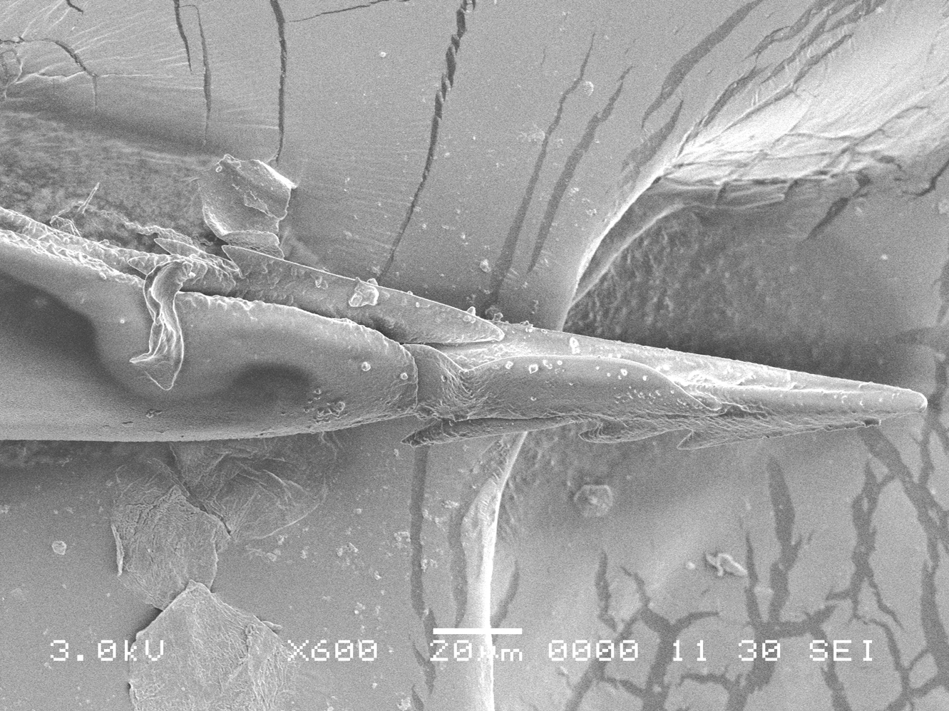
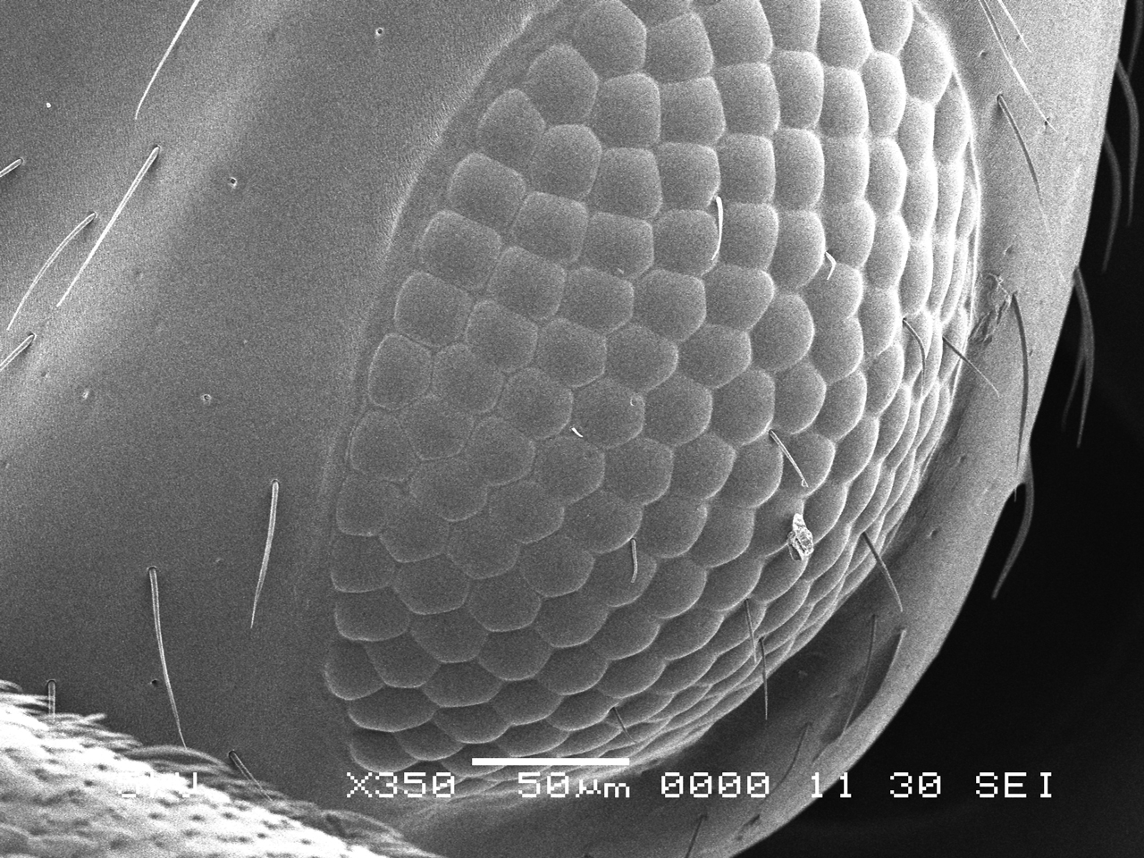
Scanning Electron Microscope
Scanning electron microscopy is an analytical technique for imaging objects in the magnification range of 25x-30,000x. An image is produced by firing a beam of electron at conductive or gold-plated specimen. Since no light is used, images are grey scale but have the umatched combination of large depth of field and high resolution. Images of different resolutions are created by differing the scan times of the samples. The photographic images here are from 120 second scans from the instrument at Florida Institute of Technology and I would like thank microscopy technician Gayle Duncombe for her help.